Small Worlds
The Nikon International Small World Competition awards photographers for their renderings of the beauty and complexity of teensy things through the light microscope. The resulting photomicrographs must not only awe but also contribute significantly to various scientific disciplines. From alien-looking insects and worms to cells that would make stunning decorations, the 2011 award-winning photographs are both stunning and informative. Take a stroll into the world of the small. [This list is according to rank (prize winner on top)].
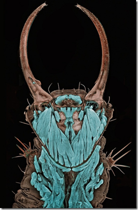
1. Tiny Monster
Photograph of an itsy-bitsy green lacewing larva.
Credit: Dr. Igor Siwanowicz Max Planck Institute of Neurobiology Martinsried, Germany.
2. Leaves of Grass
A blade of grass magnified 200 times.
Credit: Dr. Donna Stolz University of Pittsburgh Pittsburgh, Pennsylvania, USA.
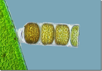
3. Bit of Algae
Photo of living Melosira monliformis, a type of algae.
Credit: Frank Fox Fachhochschule Trier Trier, Rheinland-Pfalz, Germany.
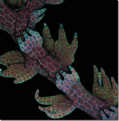
4. Fluorescent Beauty
Magnified image of liverwort.
Credit: Dr. Robin Young The University of British Columbia Vancouver, British Columbia, Canada.
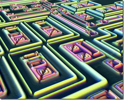
5. A-maze-ing
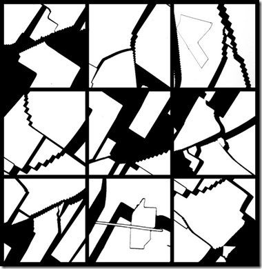
Get ultra-close with electronics: The surface of a microchip.
Credit: Alfred Pasieka Germany.
6. Broken Windows
Picture of cracked gallium arsenide solar cell films.
Credit: Dennis Callahan California Institute of Technology Pasadena, California, USA.
7. Bright Lights
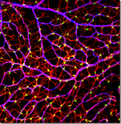
Image of mouse nerve fibers.
Credit: Gabriel Luna UC Santa Barbara, Neuroscience Research Institute Santa Barbara, California, USA.
8. Stained Glass Rocks
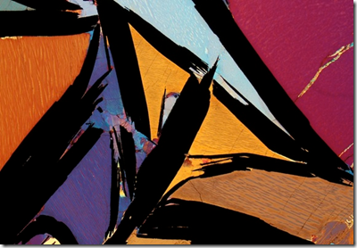
Close-up image of a graphite-bearing granulite from India.
Credit: Dr. Bernardo Cesare Department of Geosciences Padova, Italy. See more of his beautiful photography here.
9. Itsy-Bitsy Ocean-Dweller
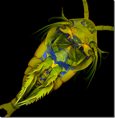
The marine copepod Temora longicornis takes on subtle hues.
Credit: Dr. Jan Michels Christian-Albrechts-Universität zu Kiel Kiel, Germany.
10. Hello, Flea
This is a freshwater water flea, Daphnia magna.
Credit: Joan Röhl Institute for Biochemistry and Biology Potsdam, Germany.
11. Ant Attack!
Eeeek! A fluorescing ant head/horror movie monster took.
Credit: Dr. Jan Michels Christian-Albrechts-Universität zu Kiel Kiel, Germany.
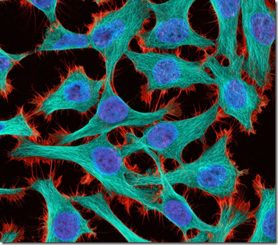
12. HeLa Cell Dance
Colourful image of cancer cells. These HeLa cells are the descendants of a seemingly immortal cancer cell line taken from the cervix of Henrietta Lacks, a black woman born in Virginia who died of her disease in 1951, never knowing how important her cells would become to medicine.
Credit: Thomas Deerinck National Center for Microscopy and Imaging Research La Jolla, California, USA.
13. Pink Triangle
A curare vine (Chondrodendron tomentosum) in cross-section.
Credit: Dr. Stephen S. Nagy Montana Diatoms Helena, Montana, USA.
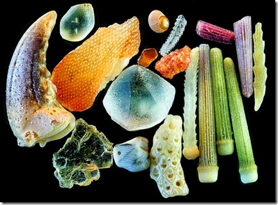
14. In a Grain of Sand
Shapely sand grains.
Credit: Yanping Wang Beijing Planetarium Beijing, China.
15. Coral Colour
A close-up look at a lobe coral's pigmentation response.
Credit: James H. Nicholson Coral Culture and Collaborative Research Facility, NOAA/NOS/NCCOS/CCEHBR & HML Charleston, South Carolina, USA.
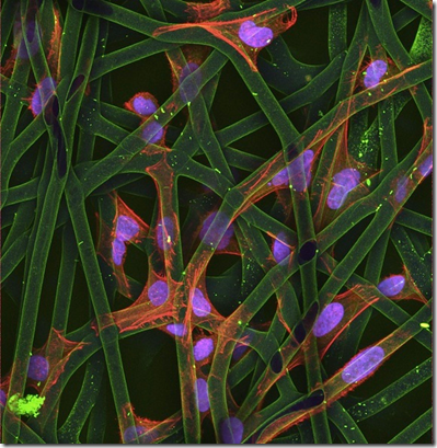
16. Scaffold of Life
Cultured cells grow on a bio-polymer scaffold.
Credit: Dr. Christopher Guérin VIB (Flanders Institute of Biotechnology) Ghent, Belgium.
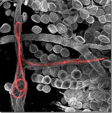
17. Earworms
Filaria worms lurk inside lymphatic vessels in a mouse's ear.
Credit: Dr. Witold Kilarski, EPFL-Laboratory of Lymphatic and Cancer, Lausanne, Switzerland.
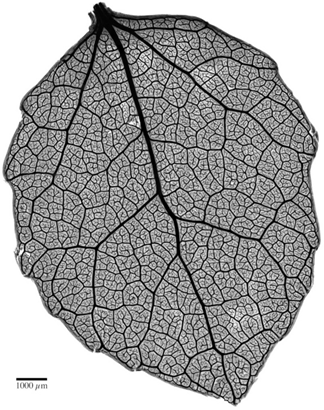
18. Quaking Lace
Delicate veins of a quaking aspen leaf, magnified four times.
Credit: Benjamin Blonder, David Elliott University of Arizona Tucson, Arizona, USA.
19. Really Tiny Christmas
A collage of mammalian cells, stained to reveal various proteins and organelles and then assembled into a wreath.
Credit: Dr. Donna Stolz The University of Pittsburgh Pittsburgh, Pennsylvania, USA.
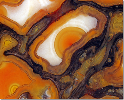
20. Dinosaur Bone Art
Image of agatized dinosaur bone cells magnified 42 times.
Credit: Douglas Moore University of Wisconsin - Stevens Point Stevens Point, Wisconsin, USA.
[Source: Live Science. Edited.]







No comments:
Post a Comment
Please adhere to proper blog etiquette when posting your comments. This blog owner will exercise his absolution discretion in allowing or rejecting any comments that are deemed seditious, defamatory, libelous, racist, vulgar, insulting, and other remarks that exhibit similar characteristics. If you insist on using anonymous comments, please write your name or other IDs at the end of your message.